HIT Consultant – Read More
What You Should Know:
– Mount Sinai-affiliated doctors in New York City have become the first in the region to integrate a validated AI software tool, BrightHeart, into large-scale clinical practice to enhance fetal ultrasounds.
– A recent study led by Mount Sinai West physicians showed that AI assistance improved the detection of suspicious findings for major congenital heart defects to over 97 percent, while simultaneously reducing reading time by 18 percent. This adoption promises to improve workflow efficiency, standardize care, and drive earlier detection of one of the most common birth abnormalities.
Mount Sinai Scales Up Fetal Diagnostics with Predictive Technology
The journey of an expectant parent is often filled with anxiety, particularly concerning fetal health. Among the most serious threats are congenital heart defects (CHDs), which affect the structure of the heart at birth. About 1 in 500 newborns is classified as having a severe CHD that requires urgent medical or surgical intervention.
Now, a major development from the Mount Sinai Health System is set to dramatically improve early detection and care coordination for these critical conditions. Doctors in the Raquel and Jaime Gilinski Department of Obstetrics, Gynecology and Reproductive Science have become the first in New York City to implement an FDA-approved artificial intelligence (AI) software tool from medical company BrightHeart on a large scale.
Carnegie Imaging for Women, a Mount Sinai-affiliated modern OB/GYN imaging facility with three Manhattan locations, is the first center in the city to deploy this AI platform, showcasing a commitment to technological innovation at the point of care.
97% Detection and Faster Workflow
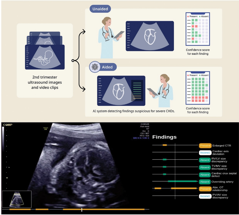
The impact of this AI integration is validated by robust quantitative data. In a recent Obstetrics & Gynecology study, Mount Sinai West researchers demonstrated a profound improvement in performance using the AI technology:
- Improved Detection: The detection rate for ultrasound findings suspicious for major congenital heart defects increased to more than 97 percent.
- Efficiency Gain: Reading time was reduced by 18 percent.
- Clinician Confidence: Physicians’ confidence scores improved by 19 percent.
The study examined a deidentified dataset of 200 fetal ultrasound examinations (18 to 24 weeks of gestation) reviewed by 14 specialists, both with and without AI assistance. The results confirmed the ability of AI-based software to improve not only the detection of suspicious lesions but also the overall confidence and time efficiency in interpreting these critical scans.
Leveling the Field in Prenatal Diagnosis
For clinicians, the benefit extends beyond raw numbers; it’s about standardization and reducing variability in care.
“AI assistance in prenatal diagnosis offers not only improved detection, but has the potential to offer significant improvement in workflow and efficiency benefits,” said corresponding author Jennifer Lam-Rachlin, MD, Assistant Clinical Professor at the Icahn School of Medicine at Mount Sinai.
Dr. Lam-Rachlin highlighted the technology’s potential to “level the field of prenatal diagnosis to offer close to expert-level review of fetal ultrasounds, particularly in centers or geographical locations without fetal heart experts”.
This sentiment was echoed by co-author Andrei Rebarber, MD, Director of the Division of Maternal-Fetal Medicine at Mount Sinai West, who noted that the study should “prompt and encourage future research into AI-assisted software’s ability to improve detection rates… to reduce the variability and inequity of detection of congenital heart defects globally”.
The integration of AI as an adjunct to physician interpretation signals a bright future for prenatal diagnostic imaging, ensuring earlier detection and better outcomes for babies and their families.